
Quality Standards
Made in America
Proudly manufactured in the USA
Third-Party Tested
Independently tested for purity and quality
>99% Purity
Exceptional purity you can trust
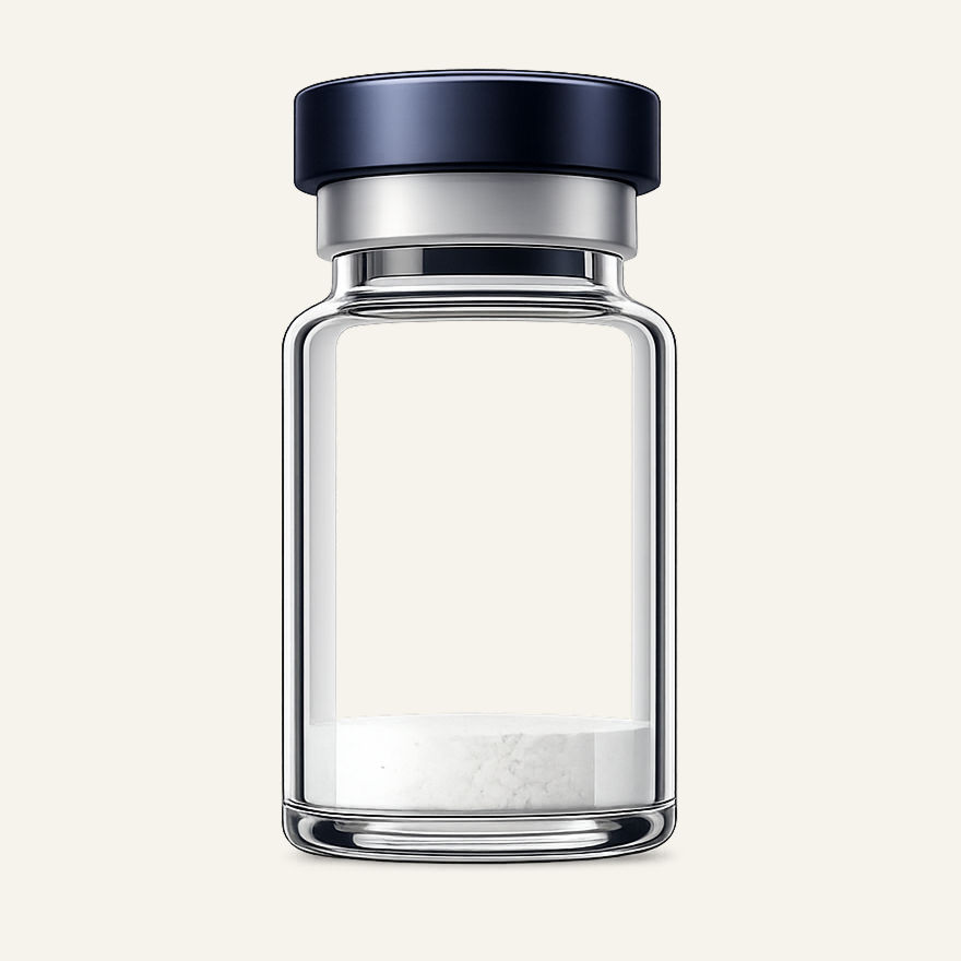
Glutathione
γ-Glutamyl-cysteinyl-glycine tripeptide intracellular redox cofactor more info
Available for laboratory research use only.
Quality Standards
Made in America
Proudly manufactured in the USA
Third-Party Tested
Independently tested for purity and quality
>99% Purity
Exceptional purity you can trust
Vial size
Choose your supply
3 vials· 4500mg total
Save $8.85 (5% off)
Total
$168.15
Volume pricing applies to your total vials, not just this product. Mix or match any vials in your cart to reach the next tier.
Independent Lab Results
The most comprehensive testing panel in research peptide commerce. Every batch is independently verified by ILS Laboratories — an ISO/IEC 17025 and PJLA-accredited facility in San Diego, CA.
- Identity
- Purity (HPLC)
- Endotoxin (USP <85>)
- Sterility (USP <71>)
- Heavy metals (ICP-MS per USP <233>)
Biochemical Profile
- CAS Number
- 70-18-8 (GSH); 27025-41-8 (GSSG)
- Molecular Formula
- C10H17N3O6S (GSH); C20H32N6O12S2 (GSSG)
- Molecular Weight
- 307.32 g/mol (GSH); 612.63 g/mol (GSSG)
- Purity
- ≥98% (HPLC-UV (210-220 nm) with Ellman's reagent (DTNB) free-thiol quantification; LC-MS at m/z 308.1 [M+H]+ (GSH) and m/z 613.2 [M+H]+ (GSSG). The free Cys-thiol is redox-sensitive; air-oxidation to GSSG is the dominant purity-failure mode and is reported as %GSSG content on lot Certificates of Analysis.)
- Amino Acid Sequence
- γ-Glu-Cys-Gly (non-canonical γ-glutamyl bond between Glu side-chain γ-carboxyl and Cys α-amine)
Intracellular Redox Cofactor and Phase-II Conjugation Substrate
Glutathione is biochemically a small-molecule redox cofactor present at 1-10 mM cytosolic concentration in mammalian cells, with the closest functional analogs being NAD(P)H and ascorbate rather than the signaling peptides typical of laboratory research catalogs[1][2]. The plasma compartment carries only 2-20 µM GSH, approximately three orders of magnitude lower than the intracellular pool, and the gradient between these two compartments is the load-bearing framing distinction for any research involving exogenous administration.
The central reactive group is the free thiol (-SH) on the cysteine residue. Glutathione peroxidases (GPx, EC 1.11.1.9), a family of selenium-dependent enzymes, catalyze the reaction in which two GSH molecules reduce one hydrogen peroxide or organic hydroperoxide, yielding one GSSG and two water molecules[1]. Glutathione reductase (GSR, EC 1.8.1.7), an NADPH-dependent flavoenzyme, regenerates two GSH from one GSSG, closing the redox cycle. NADPH for this reaction is supplied by the pentose phosphate pathway via glucose-6-phosphate dehydrogenase (G6PD); G6PD deficiency phenocopies a collapse of the GSH system in erythrocytes.
Biosynthesis is intracellular and ATP-dependent across two enzymatic steps. Glutamate-cysteine ligase (GCL, EC 6.3.2.2) conjugates glutamate to cysteine via the γ-carboxyl side chain, yielding γ-glutamylcysteine; GCL is the rate-limiting step and is feedback-inhibited by GSH itself with a Ki of approximately 2.3 mM[3]. Glutathione synthetase (GS, EC 6.3.2.3; UniProt P48637) ligates glycine onto the C-terminus of γ-glutamylcysteine, completing the tripeptide. Cysteine availability is the cellular limiting substrate, which is the biochemical basis for cysteine donors (N-acetylcysteine, γ-glutamylcysteine) producing reliable increases in intracellular GSH in published studies, whereas oral administration of intact GSH does not[4].
The second canonical biochemical role of GSH is as a substrate for the glutathione S-transferase (GST) family of phase-II conjugation enzymes. GSTs lower the pKa of the GSH thiol below 7, enabling nucleophilic addition to electrophilic carbon centers in xenobiotics and reactive metabolites of phase-I oxidation. The conjugates are exported from cells by multidrug-resistance proteins (MRPs) and excreted as mercapturic acids in urine[5]. The acetaminophen-toxicity-and-N-acetylcysteine pharmacology is the clearest documented clinical application of the phase-II conjugation system in human research.
The GSH:GSSG ratio in healthy cells is typically greater than 100:1 in cytosol; a shift toward GSSG is the textbook biomarker of cellular oxidative stress and is widely used as a readout in cell-culture redox research[6]. Approximately 10-15% of cellular GSH is in the mitochondrial matrix, where it acts as a redox buffer for the electron transport chain against reactive oxygen species; the mitochondrial pool is imported from the cytosol by specific transporters because the biosynthetic enzymes are cytosolic.
Research Applications
Cellular Redox Biochemistry
The GSH/GSSG redox couple is the dominant non-protein thiol-disulfide system in mammalian cytosol and one of the best-characterized readouts of cellular redox state in laboratory research[6]. Cytosolic concentration of reduced glutathione is 1-10 mM in most cell types and is highest in hepatocytes, where xenobiotic conjugation flux is greatest. The GSH:GSSG ratio in healthy cells is typically maintained above 100:1 by the NADPH-dependent activity of glutathione reductase, with NADPH supplied through the pentose phosphate pathway.
Cell-culture redox studies have used buthionine sulfoximine (BSO, a GCL inhibitor) to deplete cellular GSH and characterize the consequences for oxidative damage, chemotherapy sensitivity, and ferroptosis induction. Stable-isotope tracer methodology and Grx-roGFP biosensor preparations have been deployed to distinguish intracellular pools from extracellular medium contributions, since extracellular GSH oxidation in culture medium can confound naive interpretation of redox-state readouts[2].
The Sies framing of GSH as the principal intracellular thiol antioxidant has been the dominant conceptual model in cellular redox biochemistry across four decades of published research[1][6].
ROS Detoxification
Reactive oxygen species detoxification through glutathione peroxidase (GPx) is one of the canonical mechanisms by which mammalian cells handle hydrogen peroxide and organic hydroperoxides. The selenium-dependent GPx family catalyzes the reaction 2 GSH + ROOH → GSSG + ROH + H2O, using the cysteine thiol of glutathione as the nucleophilic reductant[1]. Eight GPx isoforms have been described in the human genome, with GPx1 (cytosolic) and GPx4 (membrane phospholipid hydroperoxide GPx) being the most studied in cellular and tissue research preparations.
GPx4 in particular has been investigated as the principal cellular defense against lipid peroxidation, and its inhibition or knockout is the canonical experimental induction of ferroptosis, a form of regulated cell death distinct from apoptosis. The Forman, Zhang, and Rinna review remains the standard methodological reference for measurement of GSH-dependent peroxide detoxification in cell-culture and tissue research[6].
The overall ROS detoxification network is interconnected with selenium status (required for active-site selenocysteine in GPx), NADPH availability (required for GSSG reduction), and cysteine availability (rate-limiting for GSH biosynthesis).
Phase-II Conjugation
The glutathione S-transferase (GST) enzyme family catalyzes the conjugation of GSH to electrophilic substrates as the principal phase-II xenobiotic-metabolism pathway in mammalian liver and other tissues. GSTs are present as seven main classes in humans (alpha, mu, pi, theta, zeta, omega, sigma) with overlapping but distinct substrate preferences. The mechanistic feature of GST catalysis is the lowering of the GSH-thiol pKa from approximately 9 in free solution to below 7 in the active site, generating a reactive thiolate at physiological pH[5].
The GSH conjugates produced by GST catalysis are exported from cells by multidrug-resistance-associated proteins (MRPs, ABCC family) and subsequently processed by γ-glutamyltransferase and dipeptidases to the corresponding cysteine S-conjugates, which are then N-acetylated by N-acetyltransferase 8 to form mercapturic acids that are excreted in urine. This is the textbook phase-II conjugation excretion pathway and is the basis for one of the few clinical applications of N-acetylcysteine: as the FDA-approved antidote for acetaminophen-induced hepatotoxicity, where NAC supplies cysteine for hepatic GSH biosynthesis and the resulting GSH conjugates the toxic NAPQI metabolite of acetaminophen[18].
Mitochondrial Function
Approximately 10-15% of total cellular glutathione is localized to the mitochondrial matrix, where it serves as the redox buffer for the electron transport chain against reactive oxygen species generated by complex I and complex III leak[2]. The mitochondrial GSH pool cannot synthesize its own glutathione, because the biosynthetic enzymes (GCL and GS) are cytosolic. The matrix pool is therefore imported from the cytosol by specific transporters in the inner mitochondrial membrane, including the dicarboxylate carrier and the 2-oxoglutarate carrier.
Mitochondrial GSH depletion sensitizes cells to apoptotic stimuli, mitochondrial outer membrane permeabilization (MOMP), and ferroptotic cell death. The mitochondrial-redox biology is the principal preclinical mechanistic substrate for hypotheses involving glutathione status and neurodegenerative disease, since substantia nigra mitochondrial dysfunction is a documented pathological feature of Parkinson's disease[7].
The Hauser pilot trial and the Mischley intranasal trial in Parkinson's disease research were both framed against this mitochondrial-redox rationale, although the published primary endpoints did not reach statistical significance for either route of administration in those trials[7][8].
Clinical Trial Evidence Base
The glutathione clinical-trial registry on ClinicalTrials.gov contains hundreds of NCT entries spanning oncology adjunct, Parkinson's disease, cystic fibrosis, fatty liver, autism, and dermatological cosmesis indications. The two pivotal published trials most frequently cited in the integrative-medicine literature are NCT01177319 (IV GSH in Parkinson's disease, Hauser 2009 at the University of South Florida, n=21 randomized; UPDRS difference of 2.8 units between arms with P = 0.32, not statistically significant on the primary endpoint)[7] and NCT02424708 (Phase IIb intranasal GSH in Parkinson's disease, Mischley 2017 at Bastyr University, n=45 across three arms; the principal-investigator-stated conclusion was that the data do not indicate the intervention is superior to placebo after a three-month treatment period)[8].
A precursor Phase I/IIa intranasal trial reported within-arm UPDRS observations but the subsequent Phase IIb did not reproduce those signals against placebo[15]. The broader IV-glutathione clinical evidence has produced consistent results in one narrow indication: cisplatin-chemotherapy adjunct for reduction of cisplatin-induced peripheral neuropathy and nephrotoxicity[9]. The Italian Tationil and TAD products are authorized in EU jurisdictions for this oncology-adjunct indication. Across the wider commerce categories (skin-lightening cosmesis, athletic performance, chronic Lyme infusions, anti-aging wellness drips), the published clinical-trial record does not support the marketed claims[17].
Replication & Regulatory Status
The intracellular-versus-injectable framing problem is the central methodology critique for exogenous glutathione research. Plasma GSH concentration (2-20 µM) and cytosolic GSH concentration (1-10 mM) differ by approximately three orders of magnitude; intravenous infusion can raise the plasma pool but does not necessarily raise the relevant intracellular pool in the target tissue, because cell membranes are not freely permeable to intact GSH[2][4]. The Hauser 2009 Parkinson's disease pilot (NCT01177319, P = 0.32 on UPDRS) and the Mischley 2017 Phase IIb intranasal trial (NCT02424708, not superior to placebo on the primary endpoint) are the pivotal negative trials commonly cited in commerce contexts in inverted form from their published conclusions[7][8].
The injectable-from-dietary-supplement-powder commerce is the cleanest documented patient-harm pattern in the glutathione literature. The Sydney 2018 endotoxin cluster (Johnstone 2018) reported seven cases of probable endotoxin poisoning at a complementary-medicine clinic following IV glutathione infusion, with five of the seven indicated for off-label chronic Lyme disease[10]. The Letco Medical 2019 FDA alert (February 1, 2019) reported seven adverse events including hospitalization in a US outpatient clinic following IV administration of glutathione compounded from Letco bulk powder; FDA testing found bacterial endotoxin at approximately five times the appropriate limit, and the powder had been distributed to approximately 100 compounders across 30 states[11]. The Letco powder was labeled 'Caution: Dietary Supplement' and 'For Manufacturing, Processing, Repackaging and Pharmaceutical Compounding', explicitly excluding sterile-injectable use[12].
The Philippines FDA Advisory No. 2019-182 (2019) characterized injectable glutathione for skin-lightening as unsafe, citing hepatotoxicity, nephrotoxicity, Stevens-Johnson syndrome risk, and the absence of published clinical trials demonstrating that injectable glutathione whitens skin; the Philippines FDA approves injectable glutathione only as an adjunct in cisplatin chemotherapy[13]. The Pharmacy Compounding Advisory Committee (PCAC) voted 8-5 with one abstention in June 2022 to recommend glutathione for the 503A Bulks List against FDA staff's recommendation; as of May 2026, FDA has not adopted the PCAC vote and glutathione is not on the interim or final 503A Bulks List[14][16].
Reconstitution & Storage
- Recommended Diluent
- Sterile water. The free Cys-thiol of reduced GSH oxidizes rapidly in aqueous solution exposed to air; antioxidant-protected preparations (low pH buffer, EDTA, inert-gas headspace) extend solution stability.
- Storage (lyophilized)
- -20°C, dry, dark, desiccated, inert-gas headspace; sealed amber vial. Refrigerated (2-8°C) storage acceptable for shorter durations.
- Storage (reconstituted)
- 2-8°C, antioxidant-protected, use within days. Aqueous reduced GSH oxidizes to GSSG within days to a few weeks at neutral pH and ambient oxygen exposure; the Cys-thiol redox sensitivity is the dominant solution-phase failure mode.
- Shelf Life
- 36 months lyophilized at -20°C with desiccant and inert-gas headspace; 24 months is the conventional pharmacopeial claim. 12-18 months at 4°C; 6-12 months at room temperature with humidity and oxygen acceleration of the oxidation-to-GSSG failure mode.
Research References
- [1] Sies H. Glutathione and its role in cellular functions. Free Radic Biol Med. 1999;27(9-10):916-921. PMID:10569624
- [2] Meister A, Anderson ME. Glutathione. Annu Rev Biochem. 1983;52:711-760. PMID:6137189
- [3] Lu SC. Glutathione synthesis. Biochim Biophys Acta. 2013;1830(5):3143-3153. doi:10.1016/j.bbagen.2012.09.008PMID:22995213
- [4] Aldini G, Altomare A, Baron G, Vistoli G, Carini M, Borsani L, Sergio F. N-Acetylcysteine as an antioxidant and disulphide breaking agent: the reasons why. Free Radic Res. 2018;52(7):751-762. PMID:29742944
- [5] Hayes JD, Flanagan JU, Jowsey IR. Glutathione transferases. Annu Rev Pharmacol Toxicol. 2005;45:51-88. PMID:15822171
- [6] Forman HJ, Zhang H, Rinna A. Glutathione: overview of its protective roles, measurement, and biosynthesis. Mol Aspects Med. 2009;30(1-2):1-12. PMID:18796312
- [7] Hauser RA, Lyons KE, McClain T, Carter S, Perlmutter D. Randomized, double-blind, pilot evaluation of intravenous glutathione in Parkinson's disease. Mov Disord. 2009;24(7):979-983. doi:10.1002/mds.22401PMID:19230029
- [8] Mischley LK, Lau RC, Shankland EG, Wilbur TK, Padowski JM. Phase IIb Study of Intranasal Glutathione in Parkinson's Disease. J Parkinsons Dis. 2017;7(2):289-299. PMID:28436395
- [9] Sechi G, Deledda MG, Bua G, Satta WM, Deiana GA, Pes GM, Rosati G. Reduced intravenous glutathione in the treatment of early Parkinson's disease. Prog Neuropsychopharmacol Biol Psychiatry. 1996;20(7):1159-1170. PMID:8916217
- [10] Johnstone T, Quinn E, Tobin S, Davis R, Najjar Z, Battye B, Gupta L. Seven cases of probable endotoxin poisoning related to contaminated glutathione infusions. Epidemiol Infect. 2018;146(7):931-934. doi:10.1017/S0950268818000766PMID:29655386
- [11] U.S. Food and Drug Administration. FDA warns compounders not to use glutathione from Letco Medical to compound sterile drugs. Drug Safety and Availability statement; February 1, 2019.
- [12] U.S. Food and Drug Administration. FDA highlights concerns with using dietary ingredient glutathione to compound sterile injectables. Human Drug Compounding statement; 2019.
- [13] Philippines Food and Drug Administration. FDA Advisory No. 2019-182: Unsafe Use of Glutathione as Skin Lightening Agent. Republic of the Philippines, Department of Health; 2019.
- [14] American Association of Pharmaceutical Compounders. PCAC recommends glutathione for bulks list. APC summary of the June 2022 Pharmacy Compounding Advisory Committee vote (8-5-1 recommendation against FDA staff position; FDA had not adopted as of May 2026).
- [15] Mischley LK, Leverenz JB, Lau RC, Polissar NL, Neradilek MB, Samii A, Standish LJ. A randomized, double-blind phase I/IIa study of intranasal glutathione in Parkinson's disease. Mov Disord. 2015;30(12):1696-1701. PMID:26230671
- [16] Anderson ME. On the discovery of glutathione. Trends Biochem Sci. 1988;13(11):422-425. doi:10.1016/0968-0004(88)90148-X
- [17] Mookherjee N, et al. Exploring the Safety and Efficacy of Glutathione Supplementation for Skin Lightening: A Narrative Review. 2024. PMID:39754511
- [18] Tenório MCDS, Graciliano NG, Moura FA, Oliveira ACM, Goulart MOF. N-Acetylcysteine (NAC): Impacts on Human Health. Antioxidants (Basel). 2021;10(6):967. PMID:34208683
Scientific Journal Author
Frederick Gowland Hopkins, FRS (deceased 1947)
Sir William Dunn Institute of Biochemistry, University of Cambridge (historical)
Landmark Publications
- Hopkins FG. On an autoxidisable constituent of the cell. Biochem J. 1921;15(2):286-305. (Original isolation of glutathione from yeast and animal tissue; initially characterized as a dipeptide of glutamate and cysteine.)
- Hopkins FG. On glutathione: a reinvestigation. J Biol Chem. 1929;84(1):269-320. (Redetermination of the structure as a tripeptide containing glutamate, cysteine, and glycine; documented the γ-glutamyl bond topology.)
- Anderson ME. On the discovery of glutathione. Trends Biochem Sci. 1988;13(11):422-425. (Historical account of the de Rey-Pailhade 1888 'philothion' observation and the Hopkins 1921/1929 isolation and structural redetermination.)
Sir Frederick Gowland Hopkins (1861-1947) is independently cited here as the originating researcher who isolated glutathione and characterized its tripeptide structure at the University of Cambridge between 1921 and 1929. Hopkins received the 1929 Nobel Prize in Physiology or Medicine. He is deceased; this citation references his historical role in the original peer-reviewed characterization of the molecule. There is no affiliation or commercial relationship between the Hopkins estate, the University of Cambridge, and Peerless Peptides.