
Quality Standards
Made in America
Proudly manufactured in the USA
Third-Party Tested
Independently tested for purity and quality
>99% Purity
Exceptional purity you can trust
KPV
α-MSH 11-13 tripeptide, C-terminal fragment Lys-Pro-Val more info
Available for laboratory research use only.
Quality Standards
Made in America
Proudly manufactured in the USA
Third-Party Tested
Independently tested for purity and quality
>99% Purity
Exceptional purity you can trust
Vial size
Choose your supply
3 vials· 30mg total
Save $11.85 (5% off)
Total
$225.15
Volume pricing applies to your total vials, not just this product. Mix or match any vials in your cart to reach the next tier.
Independent Lab Results
The most comprehensive testing panel in research peptide commerce. Every batch is independently verified by ILS Laboratories — an ISO/IEC 17025 and PJLA-accredited facility in San Diego, CA.
- Identity
- Purity (HPLC)
- Endotoxin (USP <85>)
- Sterility (USP <71>)
- Heavy metals (ICP-MS per USP <233>)
Biochemical Profile
- CAS Number
- 67727-97-3
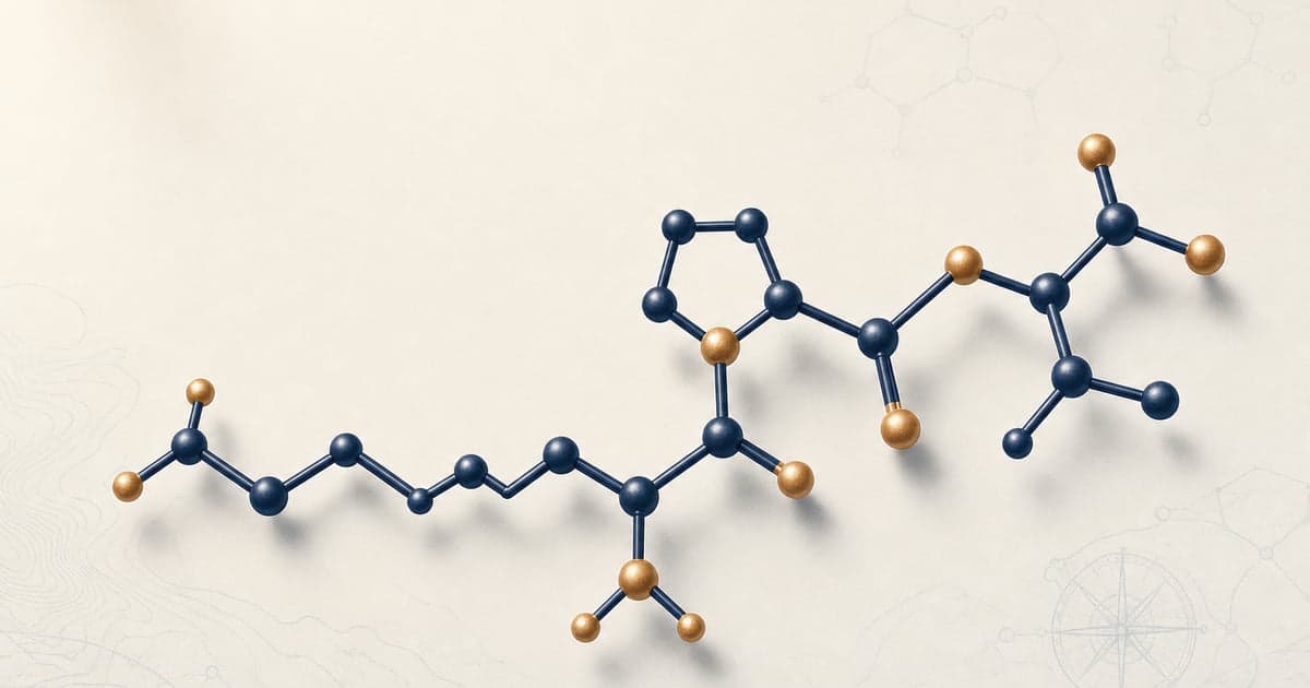
- Molecular Formula
- C16H30N4O4
- Molecular Weight
- 342.44 g/mol
- Purity
- ≥99% (HPLC-UV (210-220 nm))
- PubChem CID
- 125672
- Amino Acid Sequence
- H-Lys-Pro-Val-OH
Receptor Targets and Signaling Pathway Context
KPV has been investigated as the minimal C-terminal anti-inflammatory fragment of α-MSH. The most striking feature of the mechanism literature is that multiple primary studies have reported KPV anti-inflammatory activity that is independent of melanocortin-receptor signaling, despite KPV being derived from the prototypical melanocortin-receptor agonist α-MSH. The full parent hormone binds melanocortin receptors (MC1R, MC3R, MC4R, MC5R) via the central His-Phe-Arg-Trp (HFRW) tetrapeptide pharmacophore at residues 6-9; the C-terminal Lys-Pro-Val tripeptide does not contain that motif[1].
Getting et al. (2003) conducted a direct head-to-head comparison of core α-MSH peptides (containing the HFRW pharmacophore) against the C-terminal KPV fragment in a mouse peritonitis preparation. Core α-MSH peptides activated macrophages through melanocortin receptors and elevated cAMP; KPV failed to elevate cAMP, consistent with absence of canonical MC-R-coupled adenylate cyclase signaling, yet reduced immune-cell accumulation comparably to full α-MSH. The authors concluded that KPV is unlikely to act through melanocortin receptors and more likely acts through inhibition of IL-1β functions[2]. Brzoska, Böhm, Lügering, Loser, and Luger consolidated this position in their 2010 review, framing KPV as a fragment that lacks the entire MC-R-binding motif yet retains substantial anti-inflammatory capacity[3].
The most mechanistically explicit account of KPV's MC-receptor-independent action is Land (2012). Working in human bronchial epithelial (BEAS-2B) cells, Land reported that KPV translocated into the cell nucleus and competed for importin-α3 binding with the NF-κB p65/RelA subunit, blocking p65 nuclear import without directly affecting upstream IκBα phosphorylation. Net effect: NF-κB-driven gene transcription (IL-8, eotaxin, MMP-9) was suppressed after TNFα or rhinosyncytial-virus challenge. This is mechanistically distinct from canonical NF-κB inhibitors that act on the IκB kinase complex and distinct from melanocortin-receptor / cAMP / PKA / CREB signaling[4].
The distinctive feature of KPV pharmacology is oral bioavailability via the intestinal di/tripeptide transporter PepT1 (SLC15A1). Dalmasso et al. (2008) at Emory reported that Caco-2/BBE intestinal epithelial cells transport KPV with Km of approximately 160 micromolar for human PepT1, among the lowest Km values for hPepT1 substrates. PepT1 is normally restricted to small intestine but is inducibly expressed in inflamed colonic epithelium during inflammatory bowel disease, providing an inflammation-targeted transporter. Nanomolar intracellular KPV concentrations inhibited NF-κB activation, suppressed MAP-kinase signaling, and reduced TNFα and IL-1β secretion in intestinal epithelial and T cells[5].
The receptor-binding profile of KPV in human tissue has not been fully characterized. No direct melanocortin-receptor binding affinity has been established for the 3-residue tripeptide in radioligand assays. Some derivative vendor literature asserts MC1R / MC3R agonism for KPV, but the load-bearing primary papers (Getting 2003, Brzoska 2010, Land 2012) characterize KPV as functionally receptor-independent.
Research Applications
Gastrointestinal and Colitis Research
The strongest published preclinical case for KPV sits in murine acute colitis models induced by dextran sulfate sodium (DSS) or 2,4,6-trinitrobenzenesulfonic acid (TNBS). Dalmasso et al. (2008) at Emory reported that oral KPV administration in drinking water reduced disease activity score, attenuated colonic shortening, and lowered pro-inflammatory cytokine expression in both DSS- and TNBS-colitis preparations in mice. The same paper established the PepT1 transporter as the mechanism by which orally administered KPV reaches intestinal epithelium and immune cells at biologically active concentrations[5].
Kannengiesser et al. (2008) at the University of Münster independently reported anti-inflammatory observations for melanocortin-derived KPV in murine inflammatory bowel disease preparations, providing one of the cleaner cross-institutional replications in the KPV literature[6]. Xiao et al. (2017) at Emory extended the program through delivery optimization, reporting that hyaluronic-acid-functionalized nanoparticles (approximately 272 nm hydrodynamic diameter, slightly negative zeta potential) delivered KPV to colonic macrophages (74.7% uptake) and epithelial cells, with anti-inflammatory observations at orders-of-magnitude lower nominal peptide dose than free KPV[7].
A complicating observation: Viennois et al. (2016) at the same Emory laboratory reported that PepT1 itself plays a role in colitis-associated cancer (CAC) in murine models, even while KPV exerted anti-inflammatory effects via the same transporter. The full biological framing is not fully reconciled in the published record.
Dermatologic and Contact Dermatitis Research
The foundational dermatologic observation in the KPV literature is Hiltz and Lipton (1989), who used a picryl-chloride contact-dermatitis ear-swelling model in BALB/c mice as the dose-dependent readout for KPV anti-inflammatory activity. Graded doses of synthetic α-MSH(11-13) inhibited ear-swelling contact dermatitis in a dose-dependent fashion, identifying the C-terminal tripeptide as a candidate minimal anti-inflammatory message sequence of the parent hormone[1].
The Münster dermatology axis (Luger, Brzoska, Böhm, Loser) has expanded this body of work substantially. Brzoska et al. (2008) authored a comprehensive Endocrine Reviews piece on α-MSH and related tripeptides covering biochemistry and anti-inflammatory observations in vitro and in vivo, with particular emphasis on skin and immune-mediated inflammatory disease[8]. Luger and Brzoska (2007) framed α-MSH-related peptides as a candidate anti-inflammatory and immunomodulating class[9]. The U.S. patent estate (US 6,894,028 B2 and US Application 2002/0183255 A1) explicitly claimed method-of-use in dermatologic conditions including eczematous dermatitis. The patents have expired and the unmodified tripeptide is in the public domain[10].
A related but mechanistically distinct molecule, KdPT (Lys-D-Pro-Thr), is sometimes conflated with KPV in vendor literature. KdPT was developed by the Münster group as a D-proline-substituted analog with a different proposed mechanism (IL-1β / IL-1R signaling antagonism rather than NF-κB nuclear-import competition); Mykicki et al. (2017) reported KdPT activity in murine and human psoriasis-like skin inflammation[11].
Pulmonary and Bronchial Epithelium Research
The load-bearing pulmonary paper is Land (2012). Working in human bronchial epithelial (BEAS-2B) cells, Land reported that KPV suppressed TNFα- and rhinosyncytial-virus-induced IL-8 and eotaxin secretion. Mechanism: KPV translocated into the cell nucleus and competed for importin-α3 binding with the NF-κB p65/RelA subunit, blocking p65 nuclear import without directly affecting upstream IκBα phosphorylation[4].
The paper drew an explicit distinction between two convergent but mechanistically separate anti-inflammatory strategies for the airway. KPV acts via nuclear-import competition and is receptor-independent. γ-melanocyte-stimulating hormone (γ-MSH), the dominant MC3R agonist in airway epithelium, acts via canonical G-protein-coupled receptor signaling and elevates cAMP. The pharmacology cleanly separates the two: γ-MSH activates cAMP and behaves as an MC3R agonist; KPV does not. This is internally consistent with Getting et al. (2003), which reported no cAMP response to KPV in macrophages[2][4].
In vivo asthma-model efficacy data for KPV in murine airway-inflammation preparations are sparse in the published literature. The Land 2012 finding has not, to the present authors' knowledge, been independently replicated outside the Land laboratory in a non-BEAS-2B human bronchial cell system.
Cytokine Suppression and NF-κB Signaling Research
Across keratinocyte, intestinal epithelial, T cell, and bronchial epithelial systems, multiple groups have reported that KPV suppresses TNFα-, IL-1β-, and lipopolysaccharide-stimulated NF-κB activation. Brzoska, Luger, and the Münster axis reported attenuated p65 nuclear accumulation and reduced inflammatory cytokine output (IL-1β, IL-6, IL-8, TNFα) in human keratinocyte preparations, sometimes accompanied by IL-10 induction[8].
The Merlin group at Emory reported reduced TNFα and IL-1β secretion and reduced MAP-kinase activation in intestinal epithelial and T cells exposed to nanomolar KPV concentrations[5]. The Land 2012 paper extended the NF-κB observation to human bronchial epithelium and proposed importin-α3 competition as the upstream mechanism[4].
The convergence across three institutional axes (Münster keratinocyte, Emory intestinal, Land airway) is among the more reproducibly characterized features of the KPV literature. Whether all three observations rest on the same Land 2012 importin-α3 nuclear-import competition mechanism, or whether independent mechanisms produce similar downstream NF-κB readouts, has not been fully resolved by side-by-side mechanistic comparison.
Ocular, Peritonitis, and Fever Research
The broader α-MSH literature has reported that intravenous full α-MSH suppressed mean uveitis scores in experimental autoimmune uveitis (EAU) preparations, with effects linked to regulatory T cell induction. Luger and Brzoska (2007) reviewed this work[9]. The translation from full α-MSH (which engages melanocortin receptors and can activate Treg-relevant pathways) to KPV-only effects is less direct, since the MC-receptor pharmacophore is absent in KPV. Direct KPV-specific ocular studies are thinner than the colitis or dermatology literature.
Getting et al. (2003) used a mouse peritonitis preparation to establish the MC-receptor-independence of KPV anti-inflammatory action, with KPV reducing immune-cell accumulation comparably to full α-MSH without elevating cAMP[2].
Earlier Lipton-group work in the 1980s and 1990s used pyrogen-induced fever preparations in rodents to characterize α-MSH antipyretic activity, with KPV identified as retaining a substantial fraction of the antipyretic signal of the parent hormone. The Catania et al. (2004) Pharmacological Reviews summary catalogues this older body of melanocortin-receptor-targeting research and places KPV within the broader anti-inflammatory melanocortin program at UT Southwestern[12].
Replication, Clinical Status, and Methodology Critique
Citation network concentration on KPV is moderate. Three institutional axes dominate primary publication: the Lipton / Catania axis (UT Southwestern, then University of Milan and Weill Cornell) as the origin of the field; the Merlin / Sitaraman / Dalmasso axis at Emory and Georgia State, dominating the PepT1 transporter and oral-colitis subliterature; and the Luger / Brzoska / Böhm / Loser axis at the University of Münster, dominating the dermatology and NF-κB nuclear-translocation subliterature. A smaller fourth cluster (Land at the University of Dundee) dominates the airway sub-literature. This concentration is less extreme than BPC-157 (~87% Sikirić-affiliated) or Epitalon (~75-90% Khavinson-affiliated), but more concentrated than the literature on FDA-approved peptides.
Three methodology angles dominate the rigorous critique of the KPV evidence base. First, the full-α-MSH-to-KPV inferential bridge is overused in derivative literature. The Hiltz/Lipton 1989 origin paper[1], the Dalmasso 2008 PepT1 paper[5], and the Land 2012 importin-α3 paper[4] specifically used the 3-aa tripeptide. Many subsequent reviews and vendor copy carry full-α-MSH findings (especially ocular uveitis) into the 'KPV is anti-inflammatory' inference uncritically, despite the absence of the MC-receptor pharmacophore in KPV. Second, no peer-reviewed Phase 1, Phase 2, or Phase 3 RCT of KPV peptide for any indication has been published. ClinicalTrials.gov searches return no registered trial of KPV specifically as the investigational product[13]. Third, the PepT1 oral-bioavailability mechanism is mechanistically credible but quantitatively incomplete; published quantitative oral bioavailability data for KPV in humans are absent, and the native peptide is degraded to constituent amino acids within roughly 24 hours under pronase challenge in vitro, with Lys-Pro diketopiperazine as the major degradation product[14].
The regulatory status as of May 2026: FDA removed KPV from 503A Category 2 (Do-Not-Compound) effective April 22, 2026, as one of the 12-peptide Kennedy reclassification cohort. The Pharmacy Compounding Advisory Committee (PCAC) is scheduled to review KPV (both free-base and acetate forms) on July 23, 2026, alongside BPC-157, TB-500, and MOTS-c, with a proposed indication framed as wound healing and inflammatory conditions. Removal from Category 2 does not confer Category 1 status or compoundability[15]. KPV is not explicitly named on the WADA 2026 Prohibited List, though the S0 (Non-Approved Substances) catch-all may apply at competition control[16].
Research Literature
Published literature reviews from the Peerless research desk that reference KPV.
Reconstitution & Storage
- Recommended Diluent
- Sterile water
- Storage (lyophilized)
- -20°C, dry, dark, sealed amber vial with desiccant
- Storage (reconstituted)
- 2-8°C, use within 14-30 days
- Shelf Life
- 24+ months lyophilized at -20°C
Research References
- [1] Hiltz ME, Lipton JM. Antiinflammatory activity of a COOH-terminal fragment of the neuropeptide alpha-MSH. FASEB J. 1989;3(11):2282-2284. PMID:2550304
- [2] Getting SJ, Schiöth HB, Perretti M. Dissection of the anti-inflammatory effect of the core and C-terminal (KPV) alpha-melanocyte-stimulating hormone peptides. J Pharmacol Exp Ther. 2003;306(2):631-637. PMID:12750433
- [3] Brzoska T, Böhm M, Lügering A, Loser K, Luger TA. Terminal signal: anti-inflammatory effects of alpha-melanocyte-stimulating hormone related peptides beyond the pharmacophore. Adv Exp Med Biol. 2010;681:107-116. PMID:21222263
- [4] Land SC. Inhibition of cellular and systemic inflammation cues in human bronchial epithelial cells by melanocortin-related peptides: mechanism of KPV action and a role for MC3R agonists. Int J Physiol Pathophysiol Pharmacol. 2012;4(2):59-73. PMID:22837805
- [5] Dalmasso G, Charrier-Hisamuddin L, Nguyen HTT, Yan Y, Sitaraman S, Merlin D. PepT1-mediated tripeptide KPV uptake reduces intestinal inflammation. Gastroenterology. 2008;134(1):166-178. PMID:18061177
- [6] Kannengiesser K, Maaser C, Heidemann J, et al. Melanocortin-derived tripeptide KPV has anti-inflammatory potential in murine models of inflammatory bowel disease. Inflamm Bowel Dis. 2008;14(3):324-331. PMID:18092346
- [7] Xiao B, Xu Z, Viennois E, et al. Orally Targeted Delivery of Tripeptide KPV via Hyaluronic Acid-Functionalized Nanoparticles Efficiently Alleviates Ulcerative Colitis. Mol Ther. 2017;25(7):1628-1640. PMID:28143741
- [8] Brzoska T, Luger TA, Maaser C, Abels C, Böhm M. Alpha-melanocyte-stimulating hormone and related tripeptides: biochemistry, antiinflammatory and protective effects in vitro and in vivo, and future perspectives for the treatment of immune-mediated inflammatory diseases. Endocr Rev. 2008;29(5):581-602. PMID:18612078
- [9] Luger TA, Brzoska T. alpha-MSH related peptides: a new class of anti-inflammatory and immunomodulating drugs. Ann Rheum Dis. 2007;66 Suppl 3:iii52-iii55. PMID:17934096
- [10] US Patent 6,894,028 B2. Use of KPV tripeptide for dermatological disorders. Granted 2005; expired. Original assignee: Schering-Plough (subsequently Merck via 2009 merger). Method-of-use claims; composition-of-matter on the unmodified tripeptide is in the public domain.
- [11] Mykicki N, Klenner L, Baumann C, et al. The tripeptide KdPT ameliorates ongoing psoriasis-like skin inflammation in murine and human skin. Exp Dermatol. 2017;26(4):328-334. PMID:27376341
- [12] Catania A, Gatti S, Colombo G, Lipton JM. Targeting melanocortin receptors as a novel strategy to control inflammation. Pharmacol Rev. 2004;56(1):1-29. PMID:15001661
- [13] ClinicalTrials.gov registry search for 'KPV' as investigational product, verified 2026-05-19. No Phase 1, Phase 2, or Phase 3 trial of KPV (Lys-Pro-Val tripeptide) for any indication is identifiable as of this review. Substring false-positives exist (other trials with 'kpv' in sponsor or institution names) but no primary KPV trial is registered.
- [14] Russo R, Cassiano C, Carabetta A, Sirignano M, Riccio R, Casapullo A. Reductive glycoalkylation of the lysine ε-amine in tripeptide KPV: pronase-stable analogs and stability characterization of the native peptide. Bioorg Med Chem. 2018;26(8):1759-1768. PMC6023233. PMID:29472036
- [15] Frier Levitt analysis. FDA Removes 12 Peptides from 503A Category 2 (Do-Not-Compound List), Schedules July 23, 2026 PCAC Review. April 2026. KPV included in the 12-peptide Kennedy reclassification cohort; PCAC Day 1 docket alongside BPC-157, TB-500, and MOTS-c. Proposed indication: wound healing and inflammatory conditions.
- [16] World Anti-Doping Agency. 2026 Prohibited List. International Standard, effective January 1, 2026. KPV is not explicitly named on the S2 (Peptide Hormones) or S5 (Diuretics and Masking Agents) sections; the S0 (Non-Approved Substances) catch-all may apply at competition control. No publicly indexed Anti-Doping Rule Violation cases involving KPV are on the registry as of May 2026.
Scientific Journal Author
James M. Lipton, MD
Department of Physiology, University of Texas Southwestern Medical Center at Dallas
Landmark Publications
- Hiltz ME, Lipton JM. Antiinflammatory activity of a COOH-terminal fragment of the neuropeptide alpha-MSH. FASEB J. 1989;3(11):2282-2284. (PMID 2550304) — origin paper establishing KPV as the minimal C-terminal anti-inflammatory fragment of α-MSH; co-discoverer Marlene E. Hiltz.
- Catania A, Gatti S, Colombo G, Lipton JM. Targeting melanocortin receptors as a novel strategy to control inflammation. Pharmacol Rev. 2004;56(1):1-29. (PMID 15001661) — secondary-literature consolidation of the Lipton-Catania anti-inflammatory melanocortin program.
- Lipton JM, Catania A. Anti-inflammatory actions of the neuroimmunomodulator alpha-MSH. Immunol Today. 1997;18(3):140-145. (PMID 9078687) — review framing C-terminal fragments of α-MSH as candidate anti-inflammatory peptides.
Dr. Lipton is independently cited here as the originating researcher of KPV (with co-discoverer Marlene E. Hiltz) at UT Southwestern, where the Hiltz/Lipton 1989 FASEB Journal paper first characterized the C-terminal α-MSH tripeptide as a minimal anti-inflammatory fragment. There is no affiliation or commercial relationship between Dr. Lipton, UT Southwestern Medical Center, or any associated entity, and Peerless Peptides.