

Quality Standards
American Owned
Mixed, bottled, and tested in the USA
Third-Party Tested
Independently tested for purity and quality
>99% Purity
Exceptional purity you can trust
Also Screened For
Endotoxins + Heavy Metals
Beyond the identity, purity & sterility panel.
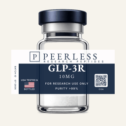
GLP-3R
Synthetic 39-amino-acid triple agonist at the GLP-1, GIP and glucagon receptors more info
Available for laboratory research use only.
Quality Standards
American Owned
Mixed, bottled, and tested in the USA
Third-Party Tested
Independently tested for purity and quality
>99% Purity
Exceptional purity you can trust
Also Screened For
Endotoxins + Heavy Metals
Beyond the identity, purity & sterility panel.
Vial size
Choose your supply
3 vials· 30mg total
Save $18.75 (5% off)
Total
$356.25
Volume pricing applies to your total vials, not just this product. Mix or match any vials in your cart to reach the next tier.
Independent Lab Results
The most comprehensive testing panel in research peptide commerce. Every batch is independently verified by ILS Laboratories — an ISO/IEC 17025 and PJLA-accredited facility in San Diego, CA.
- Identity
- Purity (HPLC)
- Endotoxin (USP <85>)
- Sterility screen (PCR)
- Heavy metals (ICP-MS per USP <233>)
Biochemical Profile
- CAS Number
- 2381089-83-2
- Molecular Formula
- C221H342N46O68
- Molecular Weight
- 4731.4 g/mol
- Purity
- >99% (HPLC-UV (214 nm))
- Amino Acid Sequence
- 39-residue GIP-derived backbone; non-coded Aib at positions 2 and 20; (αS)-α-methyl-Leu at position 13; γGlu-AEEA-C20 diacid lipidation at Lys-17 ε-amine; C-terminal serinamide
Receptor Targets and Structure
GLP-3R is a unimolecular triple agonist: one 39-residue peptide chain with agonist activity at the GLP-1 receptor (GLP-1R), the GIP receptor (GIPR) and the glucagon receptor (GCGR). In the first published pharmacological characterization, cAMP accumulation assays in cells expressing the human receptors gave EC₅₀ values of approximately 0.064 nM at GIPR, 0.775 nM at GLP-1R and 5.79 nM at GCGR[1]. Expressed against the native ligands in the same system, that is about 8.9 times the potency of native GIP at GIPR, and about 0.4 and 0.3 times the potency of native GLP-1 and native glucagon at their own receptors: balanced activity at GLP-1R and GCGR, with GIPR activity above that of the native hormone[1].
Cryo-electron microscopy structures of the peptide bound to each receptor in complex with the Gs protein were reported by Li and colleagues at overall resolutions of 2.68 Å (GLP-1R), 3.26 Å (GIPR) and 2.84 Å (GCGR), with coordinates deposited in the Protein Data Bank as 8YW3, 8YW4 and 8YW5[2]. In all three complexes the peptide forms a single continuous helix. Its N-terminal segment (residues 1-13) penetrates the core of the receptor transmembrane domain, and its C-terminal segment (residues 14-30) contacts the N-terminal helix of the receptor extracellular domain, the extracellular tip of transmembrane helix 1 and extracellular loop 1[2].
All three receptors belong to the secretin family of class B G-protein-coupled receptors and couple mainly to Gαs and cyclic AMP signaling[3]. The endogenous ligands of two of them, GLP-1 and glucagon, are both cut from proglucagon, the product of the GCG gene, by tissue-specific prohormone convertases: PC1/3 in intestinal L cells and PC2 in pancreatic α cells[3]. GIP is encoded by a separate gene and secreted by intestinal K cells[4].
The backbone is GIP-derived and carries three non-coded residues: α-aminoisobutyric acid (Aib) at positions 2 and 20 and α-methyl-leucine at position 13. The lysine at position 17 carries a C20 fatty diacid through a γGlu-AEEA linker, a lipidation motif that binds serum albumin, and the C-terminus is amidated (serinamide).
Research Applications
Triple Receptor Agonist Pharmacology Research
Receptor-binding and signaling studies characterize GLP-3R as a single peptide that engages GLP-1R, GIPR and GCGR at the same time[1]. Its potency ordering in cAMP assays (GIPR, then GLP-1R, then GCGR) makes it a model compound for imbalanced multi-receptor agonism, in which potencies at the individual receptors are set at unequal ratios to shape the integrated cellular response. The ratio of receptor potencies is the central design variable across unimolecular agonists at the GIP, GLP-1 and glucagon receptors, a peptide class reviewed by Knerr and colleagues[5].
The cryo-electron microscopy structures show how one sequence is accommodated by three receptors[2]. The three complexes are highly similar overall, with Cα root mean square deviations of 0.88 to 0.93 Å. The peptide keeps a common set of contacts with conserved receptor residues, including two salt bridges formed by the N-terminal amine of Tyr1, while receptor-specific contacts in the upper half of the transmembrane pocket absorb the sequence differences between the receptors[2]. Extracellular loop 1 forms a short α-helix in GLP-1R and GCGR but an unwound loop in GIPR, attributed to three proline residues (P195, P197 and P199). In the GIPR complex the peptide straightens, and the helix at Leu27 shifts about 4 Å toward the transmembrane core relative to the other two complexes[2].
Alanine mutagenesis in the same study weighed the shared contacts receptor by receptor. Substitution at the conserved E6.53b position lowered cAMP potency 3.6-fold at GLP-1R, 1.6-fold at GIPR and 8.3-fold at GCGR, and substitution at E/D7.42b lowered it 71.6-fold at GIPR and 5.8-fold at GCGR[2]. The coordinates are public, so the contact map is open to re-analysis by any structural biology group.
Long-Acting Acylated Peptide Engineering Research
GLP-3R is studied in the peptide chemistry literature as a model compound for γGlu-AEEA-C20 fatty diacid acylation at a lysine ε-amine, here Lys-17[1]. From the lysine outward, the arm runs through a γ-L-glutamic acid spacer and a single 8-amino-3,6-dioxaoctanoic acid (AEEA) unit to a C20 α,ω-eicosanedioic diacid, the moiety that binds serum albumin. Reversible albumin binding is the basis of the extended plasma residency of acylated peptides at the incretin receptors[3].
Three non-coded residues shape stability and conformation: Aib at positions 2 and 20 and (αS)-α-methyl-leucine at position 13[1]. Aib at position 2 removes the N-terminal dipeptidyl peptidase IV cleavage site, and the α,α-disubstituted residues at positions 13 and 20 favor the helical conformation seen along the full receptor-bound peptide in the cryo-electron microscopy structures[2]. Couplings onto and from α,α-disubstituted residues are sterically hindered steps in solid-phase synthesis.
The molecular formula contains no sulfur, so the sequence carries no methionine or cysteine and is not exposed to sulfur-oxidation chemistry. The two aspartate residues, Asp-9 and Asp-15, are the positions at which aspartimide formation, a common side reaction in Fmoc solid-phase synthesis, can occur. Hydrolysis within the lipidation arm is the other principal chemical liability.
The analytical consequence follows from the structure. Receptor activity assays measure signaling, not mass, so they do not by themselves establish that the lipidation arm is intact. Identity at the level of the arm is read by liquid chromatography mass spectrometry with deconvolution, which separates the intact parent from variants missing the C20 diacid, the AEEA unit or the γGlu spacer.
Glucagon Receptor and Hepatic Metabolism Research
The glucagon-receptor arm of GLP-3R is studied as a hepatic signaling axis distinct from the two incretin receptors. GCGR is expressed at high density on hepatocytes. In the broader endocrinology literature, hepatocyte GCGR signaling through Gαs, cAMP and protein kinase A is characterized as activating glycogen phosphorylase and glycogenolysis and inducing gluconeogenic gene expression, and in rodent and isolated-hepatocyte preparations it has been associated with mitochondrial β-oxidation of fatty acids, induction of fibroblast growth factor 21 (FGF21) and lower de novo lipogenesis.
The question specific to unimolecular co-agonism is how the receptor arms interact. GCGR signaling raises hepatic glucose output, while GLP-1R signaling in islet β cells is glucose-dependent and insulinotropic. In the preclinical systems in which GLP-3R was first characterized, GCGR activity was set at about 0.3 times that of native glucagon, and concurrent incretin-receptor activity was described as counterbalancing the glycogenolytic signal of the GCGR arm[1].
The GCGR complex also differs structurally from the other two[2]. The extracellular half of transmembrane helix 7 in GCGR shifts outward by about 4.6 Å and 3.0 Å relative to the GLP-1R and GIPR complexes, reorienting the N-terminus of the peptide by about 2.8 Å and 2.6 Å. GLP-1R and GIPR each form two hydrogen bonds with Thr7 and Ser11 of the peptide that GCGR does not, because the equivalent GCGR positions (I194 and S297) cannot form them[2]. These receptor-specific features make the GCGR arm a structurally defined object of study in its own right.
Handling & Storage
- Storage (lyophilized)
- -20°C, dry, dark, 24 months
- Storage (in solution)
- 2-8°C, use within 28 days
- Shelf Life
- 24 months lyophilized
Research References
- [1] Coskun T, et al. Cell Metab. 2022;34(9):1234-1247.e9. doi:10.1016/j.cmet.2022.07.013PMID:35985340
- [2] Li W, et al. Cell Discov. 2024;10(1):77. doi:10.1038/s41421-024-00700-0PMID:39019866
- [3] Müller TD, et al. Mol Metab. 2019;30:72-130. doi:10.1016/j.molmet.2019.09.010PMID:31767182
- [4] Boer GA, Holst JJ. Biology (Basel). 2020;9(12):473. doi:10.3390/biology9120473PMID:33339298
- [5] Knerr PJ, et al. Peptides. 2020;125:170225. doi:10.1016/j.peptides.2019.170225PMID:31786282